Electron Microscopy - Samples in Suspension
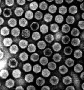
The majority of biological specimen suspension samples are examined using the electron microscopy technique of negative staining. Negative staining electron microscopy is a specialized  assay that permits the visualization and microscopic examination of various types of samples in suspension.
This unique method of sample preparation and examination allows investigators to visualize individual virus particles, proteins, peptides, bacteria cells, and other biological specimens as they actually appear in suspension. By visualizing these specimens, investigators can obtain invaluable information regarding the integrity, size, distribution, viability and morphology of their samples, and record these observations and data by means of high resolution photography.
Virtually any type of biological specimens in suspension can be examined and photographed using this technique, including:
| Virus particles | Bacteria Cells |
| Bacteriophages | Elementary Bodies |
| Lipoprotein samples | Protein particles |
| Mycoplasma cells | Cell fragments |
| Vaccine carriers | Manufactured VLP’s |
| Peptides | Protein particulates |
Service includes:
Complete sample preparation, fixation, and staining; extensive microscopic examination; six to eight 8” x 10” electron micrographs, a CD containing the digital images, and a full, detailed report describing the final sample results and material and methods.
This email address is being protected from spambots. You need JavaScript enabled to view it.


